导语:


图1. PIT-1 参与垂体前叶细胞分化

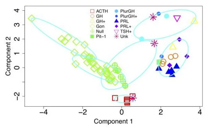
根据2017版WHO新分类,PIT-1阳性谱系腺瘤包括生长激素细胞腺瘤、泌乳激素细胞腺瘤、促甲状腺激素细胞腺瘤、PIT-1阳性激素阴性腺瘤和多激素PIT-1阳性腺瘤(以前称为静止性第三亚型腺瘤)。
生长激素腺瘤
泌乳素腺瘤

促甲状腺激素腺瘤
PIT-1阳性激素阴性腺瘤
PIT-1阳性激素阴性腺瘤表达PIT-1但不表达GH、PRL或TSH,它们可能导致GH、PRL或TSH或其中两种激素的功能亢进,应被指定为(低分化)PIT阳性、激素阴性的腺瘤。
多激素PIT-1阳性腺瘤腺瘤
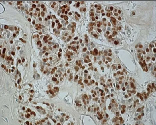
多激素PIT-1阳性腺瘤(静止性第三亚型腺瘤)侵袭性高、易复发、无病生存率低。John P. Andrews等人对Medline和Google Scholarzhong 1990-2020的病例进行了荟萃分析,并回顾了2012-2019年在自己所在中心手术治疗的腺瘤,结果表明该肿瘤的病理特征为:所有肿瘤都表现出单形性特征,转录因子PIT-1呈核弥漫强阳性表达,腺瘤细胞不同程度表达GH、PRL、TSH和α亚基,但不表达SF-1和T-PIT,如图4所示。

图4. 多激素PIT-1阳性腺瘤免疫组化特征
A: 苏木精伊红染色;B: 转录因子PIT-1弥漫强阳性;C-E:GH、PRL、TSH染色
迈新相关抗体
抗体名称 | 产品编号 | 克隆号 | 细胞定位 |
PIT-1 | MAB-0902 | MX106 | 胞核 |
参考文献:
[1]Kobalka P J , Kristin H , Becker A P . Neuropathology of Pituitary Adenomas and Sellar Lesions[J]. Neurosurgery, 2021.
[2]Mcdonald W C , Banerji N , Mcdonald K N , et al. Steroidogenic Factor 1, Pit-1, and Adrenocorticotropic Hormone: A Rational Starting Place for the Immunohistochemical Characterization of Pituitary Adenoma[J]. Archives of Pathology & Laboratory Medicine, 2017, 141(1):104-112.
[3]Lopes, Bs M . The 2017 World Health Organization classification of tumors of the pituitary gland: a summary[J]. Acta Neuropathologica, 2017.
[4]Saeger W , Koch A . Pathology of pituitary adenoma[J]. Pituitary Tumors, 2021,(26):373-392
[5]Burcea I F , Nstase V N , Poian C . Pituitary transcription factors in the immunohistochemical and molecular diagnosis of pituitary tumours - a systematic review[J]. Endokrynologia Polska, 2021, 72(1):53-63.
[6]Andrews J P , Joshi R S , Pereira M P , et al. Plurihormonal Pit-1 Positive Pituitary Adenomas: a systematic review and single center series[J]. World Neurosurgery, 2021:E1-E7.
| 上一篇什么是伴随诊断,你知道... | >>返回 | 下一篇肿瘤漫谈——毁掉一个胃... |